Description
Collection of 8 single images:
- Vestibular view of the anterior maxillary sextant. A marked horizontal soft tissue deficiency appears.
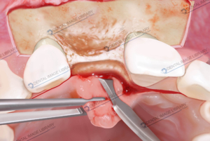
- Buccal flap design. A 45 degrees internal beveled incision, with an apical-coronal direction, with a 15c scalpel blade, is performed.
- The incision is “reinforced” to the underlying bony structure, using a blunt tip dissector.
- A small CTGO bone chisel is used to raise a full-thickness buccal flap.
- Palatal flap design. The incision extends to the base of the 2 adjacent palatal papillae. No vertical release incisions are utilized.
- The palatal incision is “enhanced”, reaching the underlying bone structure, using a blunt tip dissector.
- A small CTGO bone chisel is used to raise a full-thickness palatal flap.
- Occlusal view of the anterior maxillary sextant. A marked horizontal bone deficiency appears.
Every single image is a jpeg file:
3872×2592 pixels
32,78×21,95 cm 300 dpi


 Elisa Botton
 Elisa Botton
 Elisa Botton
 Elisa Botton
 Elisa Botton
Reviews
There are no reviews yet.