
Description
Guided Bone Regeneration – Dental Implant Bone Defect Illustration
This detailed medical illustration depicts a vestibular bone defect around a dental implant, shown in a sagittal section of the mandible. The image illustrates a guided bone regeneration (GBR) procedure used to treat defects like peri-implantitis—a condition involving bone loss around a dental implant—using two types of Bio-Oss bone graft material and a resorbable membrane. It has been created to support education and communication in implant dentistry and periodontology.
The image shows the bone defect clearly, along with the step-by-step regenerative treatment applied to correct it. The use of both granular and block-form Bio-Oss highlights different approaches within the same procedure, offering versatility for various clinical contexts. The illustration also features the precise placement of the resorbable membrane, an essential component for ensuring successful bone regeneration, especially in cases of peri-implantitis.
This visual resource is especially valuable for professionals and students in:
-
Implantology and periodontology
-
Dental and medical education
-
Academic presentations and lectures
-
Patient consultation and treatment explanation
It helps educators explain complex surgical concepts, aids clinicians in communicating procedures to patients, and supports students in understanding the steps involved in guided bone regeneration and the treatment of peri-implantitis.
Designed with scientific accuracy and visual clarity in mind, this illustration simplifies complex concepts without sacrificing anatomical detail. It was created by a professional medical illustrator with experience in dental visualization, ensuring both clinical relevance and aesthetic precision.
Key uses include:
-
Teaching materials for universities and dental schools
-
Conference posters and research presentations
-
Informational brochures and patient education
-
Online courses or continuing education programs
Technical details:
-
Format: JPEG
-
Dimensions: 1654 x 1772 pixels
-
Print size: 14 x 15 cm (300 dpi)
This image is part of the Dental Image Library collection, which features a range of high-quality visuals designed to meet the needs of modern dental professionals and educators. Whether you are preparing a course, publishing a case study, or enhancing patient understanding, this illustration offers a clear and reliable tool to support your work.
Download this image today to enrich your materials with a powerful, easy-to-understand visual aid that reflects real clinical practices in guided bone regeneration and the treatment of peri-implantitis.
 Elisa Botton
 Elisa Botton
 Elisa Botton
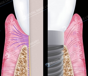 Elisa Botton
 Elisa Botton
Reviews
There are no reviews yet.