Be the first to review “Healing of the extraction socket. 102JB00041” Cancel reply
Healing of the extraction socket. 102JB00041
70,00€
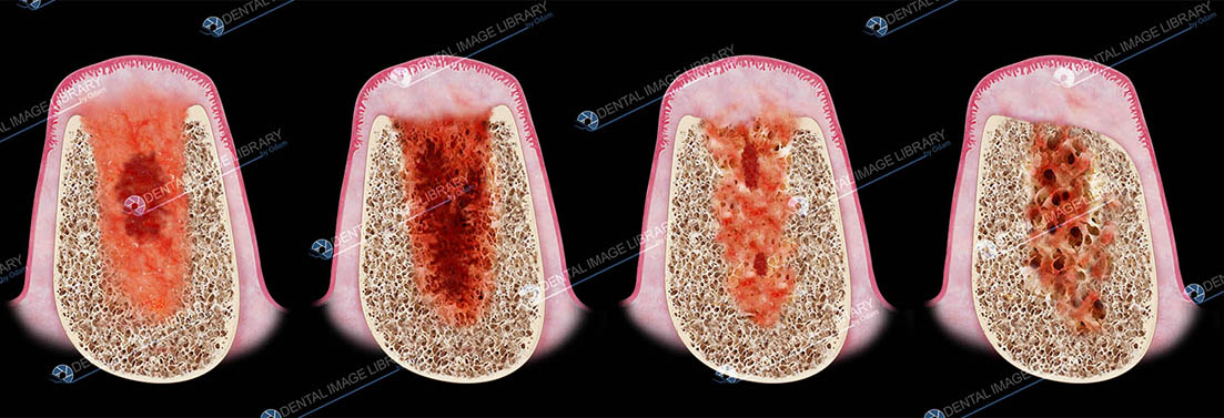
Healing of the extraction socket.
- The illustration shows a healing socket after one week. Large amounts of provisional matrix are present within the buccal and lingual wall of the sockets. And, in the center of the socket, a residual blood clot is still present.
- At two weeks the provisional matrix is still present in the most coronal and in the central part of the socket. Woven bone is rapidly forming and is already filling the apical portion of the socket. A layer of woven bone is also covering the lingual and buccal walls.
- At four weeks the socket is almost completely filled with woven bone. The white arrow is pointing an the most coronal portion of the buccal plate. In this area the bundle bone has been resorbed and replaced by woven bone.
- At eight weeks the entrance of the socket is sealed by a hard tissue that is comprised of woven bone and lamellar bone. The central portion of the socket is dominated by bone marrow. The white arrow points at the most coronal portion of the buccal plate.
Black background.
4488 x 1535 pixels
38 x 13 cm (300 dpi)
JPEG
Product added!
Browse Wishlist
The product is already in the wishlist!
Browse Wishlist
Category: Periodontology
Tags: bone loss, healing socket, Periodontology, socket, tooth socket, woven bone


 Elisa Botton
 Elisa Botton
 Elisa Botton

Reviews
There are no reviews yet.