Dental Image Library
  • Home
  • Images
    • Anatomy
    • Prosthetic dentistry
    • Dental Hygiene
    • Endodontics
    • Gnathology
    • Implantology
    • Maxillofacial Surgery
    • Oral Surgery
    • Orthodontics
    • Periodontology
  • About
  • Contact
  • Wishlist
  • Log in
  • Free Dental Illustrations
  • Menu Menu
  • 0Shopping Cart
  • Facebook
  • Instagram

SEARCH FOR IMAGES

×
  • View cart You cannot add another "Sinus floor elevation - Sinus lift. Collection of 4 illustrations. Black background" to your cart.
  • View cart You cannot add another "Inferior premolar section with periodontal disease. 102JB00076" to your cart.
View cart “One wall intrabony defect. 102JB00052” has been added to your cart.
View cart “Tissue biotype. 11JW00049” has been added to your cart.
View cart “Mandibular premolar surrounded by a healthy periodontium whit reduced height following application of traumatic occlusal forces_102JB00028” has been added to your cart.
View cart “Collection of four images: partial-thickness flaps (mucosal) and flap full-thickness (mucoperiosteal)” has been added to your cart.
View cart “Tooth with periodontal disease and inflammatory infiltrate. 102JW00049” has been added to your cart.
View cart “Sinus floor elevation – Sinus lift. Collection of 4 illustrations. Black background” has been added to your cart.
View cart “Types of bone defects. Black background” has been added to your cart.
View cart “Sagittal section of maxilla with dental implant. 61JB00058” has been added to your cart.
View cart “Inferior premolar section with periodontal disease. 102JW00072” has been added to your cart.
View cart “Tooth with healthy periodontal tissues following application of traumatic occlusal forces. 102JB00032” has been added to your cart.
View cart “Inferior premolar section with periodontal disease. 102JB00076” has been added to your cart.
View cart “Gingival recession around dental implant. 62JW00066” has been added to your cart.
View cart “Healing of the extraction socket. 102JW00040” has been added to your cart.
View cart “Collection of 3 illustrations of Guided Tissue Regeneration GTR” has been added to your cart.
View cart “Sagittal section of lower jaw with posterior edentulous ridge. 102JB00036” has been added to your cart.
View cart “The gum is then closed over reshaped bone below the original gumline. 102JW00075” has been added to your cart.
View cart “The gum is then closed over reshaped bone below the original gumline. 102JB00079” has been added to your cart.
View cart “Harvesting Epithelial-Connective tissue graft from palate. Collection of 8 images. Black background.” has been added to your cart.
View cart “The diode laser fiber inserted subgingivally. 61JB00046” has been added to your cart.
View cart “Tooth with reduced height of healthy periodontal tissues. 102JB00047” has been added to your cart.
View cart “Modified roll flap technique step by step. White background.” has been added to your cart.
View cart “The diode laser fiber inserted subgingivally. 61JW00045” has been added to your cart.
View cart “Harvesting Epithelial-Connective tissue graft from palate. Collection of 8 images. White background.” has been added to your cart.
View cart “Bone quality. 13JB00015” has been added to your cart.

The chisel of increasing width is used to split the alveolar ridge. 61JB00057

35,00€

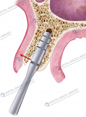
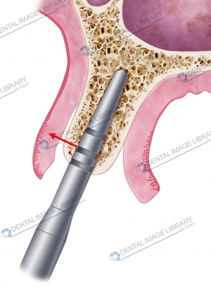
Sagittal section of maxilla with knife-edge alveolar bone morphology.

The chisel of increasing width is used to split the alveolar ridge.

Black background.

1654 x 2244 pixels

14 x 19 cm (300 dpi)

JPEG

Add to Wishlist loading
Product added! Browse Wishlist
The product is already in the wishlist! Browse Wishlist
Category: Periodontology Tags: Bone augmentation, expansion technique, horizontal augmentation, horizontal bone defect, maxilla, Ridge splitting technique, sagittal section, split crest, split osteotomy, split ridge technique
  • Reviews (0)

Reviews

There are no reviews yet.

Be the first to review “The chisel of increasing width is used to split the alveolar ridge. 61JB00057” Cancel reply

Your email address will not be published. Required fields are marked *

You may also like…

  • Ridge Split technique illustrated step-by-step. Collection of 6 illustrations. Bone augmentation

    80,00€
    Add to cart Show Details
  • The chisel of increasing width is used to split the alveolar ridge. 61JW00063

    35,00€
    Add to cart Show Details
  • The chisel of increasing width is used to split the alveolar ridge. 61JW00062

    35,00€
    Add to cart Show Details

Related products

  • Elisa Botton

    Positioning of PDS II suture material. 101JN00020

    45,00€
    Add to cart Show Details
  • Elisa Botton

    101JN00022

    45,00€
    Add to cart Show Details
  • Elisa Botton

    Bone quality. 13JW00015

    20,00€
    Add to cart Show Details
  • Elisa Botton

    101JB00007

    45,00€
    Add to cart Show Details

TERMS AND CONDITIONS – LICENSING WITH THE DENTAL IMAGE LIBRARY

© Copyright 2023 - Dental Image Library
Elisa Botton - Via Mafalda di Savoia, 24/2. 45100 Rovigo - privacy@dentalimagelibrary.com - 3280657100 - bottonelisa@pec.it
  • Facebook
  • Instagram
The chisel of increasing width is used to split the alveolar ridge. 61JB000... Sagittal section of maxilla with dental implant. 61JB00058
Scroll to top