Dental Image Library
  • Home
  • Images
    • Anatomy
    • Prosthetic dentistry
    • Dental Hygiene
    • Endodontics
    • Gnathology
    • Implantology
    • Maxillofacial Surgery
    • Oral Surgery
    • Orthodontics
    • Periodontology
  • About
  • Contact
  • Wishlist
  • Log in
  • Free Dental Illustrations
  • Menu Menu
  • 0Shopping Cart
  • Facebook
  • Instagram

SEARCH FOR IMAGES

×
View cart “Implant attachment to surrounding tissue. 62JW00039” has been added to your cart.
View cart “Gingival recession around dental implant. 62JB00067” has been added to your cart.
View cart “Guided implant surgery. 61JB00116” has been added to your cart.
View cart “All on four. 62JW00110” has been added to your cart.
View cart “Corticotomy with vertical and horizontal cuts in maxilla. 61JW00079” has been added to your cart.
View cart “Collection step-by-step illustrations. Flap passivation techniques” has been added to your cart.
View cart “Modified roll flap technique step by step. Black background.” has been added to your cart.
View cart “Peri-implant gingival fibers. 62JB00012” has been added to your cart.
View cart “Perimplantitis. 102JW00069” has been added to your cart.
View cart “Drawing of a peri-implant site with mucositis. 61JB00089” has been added to your cart.
View cart “Single screw-retained crown. 62JW00106” has been added to your cart.
View cart “Representation of tooth and implant attachment to surrounding tissue. 62JW00008” has been added to your cart.
View cart “101JB00007” has been added to your cart.

Collection of four images: partial-thickness flaps (mucosal) and flap full-thickness (mucoperiosteal)

60,00€

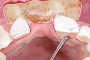
The flaps are classified as partial-thickness (mucosal) flaps and either full-thickness (mucoperiosteal).

Schematic representation of the internal bevel incision to reflect a partial-thickness flap. Note that the incision ends on the root surface to preserve the periosteum on the bone.

Schematic representation of the internal bevel incision to reflect a full-thickness (mucoperiosteal) flap. Note that the incision ends on the bone to allow for the reflection of the entire flap.

White background.

Every file is a jpeg:

3071 x 2244 pixels

26 x 19 cm

300 dpi

Add to Wishlist loading
Product added! Browse Wishlist
The product is already in the wishlist! Browse Wishlist
Categories: Oral Surgery, Periodontology, Periodontology Collections Tags: flap, flaps, full-thickness, incision, internal bevel incision, mucoperiosteal, mucosal flap, partial-thickness
  • Reviews (0)

Reviews

There are no reviews yet.

Be the first to review “Collection of four images: partial-thickness flaps (mucosal) and flap full-thickness (mucoperiosteal)” Cancel reply

Your email address will not be published. Required fields are marked *

You may also like…

  • Collection of four images: partial-thickness flaps (mucosal) and flap full-thickness (mucoperiosteal)

    60,00€
    Add to cart Show Details

Related products

  • Elisa Botton

    101JN00010

    45,00€
    Add to cart Show Details
  • Elisa Botton

    101JN00015

    45,00€
    Add to cart Show Details
  • Classification of the host bone by Lekholm and Zarb. 103JW00002

    35,00€
    Add to cart Show Details
  • Elisa Botton

    Classification of the host bone by Lekholm and Zarb. 103JB00003

    35,00€
    Add to cart Show Details

TERMS AND CONDITIONS – LICENSING WITH THE DENTAL IMAGE LIBRARY

© Copyright 2023 - Dental Image Library
Elisa Botton - Via Mafalda di Savoia, 24/2. 45100 Rovigo - privacy@dentalimagelibrary.com - 3280657100 - bottonelisa@pec.it
  • Facebook
  • Instagram
Collection of two drawings: teeth gingival fibers vs peri-implant gingival ... Collection of four images: partial-thickness flaps (mucosal) and flap full-thickness...
Scroll to top