
Description
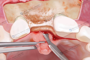
This detailed schematic illustration shows a peri-implant site affected by a peri-implant lesion, characterized by bone loss around the dental implant and the presence of an inflammatory infiltrate that may extend to the crestal bone. The image clearly highlights the progression of peri-implant disease and its potential severity.
Due to the extent of inflammation and bone involvement, the lesion can be classified as a form of osteomyelitis, sharing key features of this serious bone infection. This visual aid is ideal for clinicians, educators, and students seeking a clear and professional depiction of peri-implant pathology, useful for diagnostic explanation, patient communication, and educational materials.
 Elisa Botton
 Elisa Botton
 Elisa Botton
 Elisa Botton

Reviews
There are no reviews yet.