Dental Image Library
  • Home
  • Images
    • Anatomy
    • Prosthetic dentistry
    • Dental Hygiene
    • Endodontics
    • Gnathology
    • Implantology
    • Maxillofacial Surgery
    • Oral Surgery
    • Orthodontics
    • Periodontology
  • About
  • Contact
  • Wishlist
  • Log in
  • Free Dental Illustrations
  • Menu Menu
  • 0Shopping Cart
  • Facebook
  • Instagram

SEARCH FOR IMAGES

×
View cart “Maxillary sinus anatomy. Frontal section of the nasal cavity and sinus” has been added to your cart.
View cart “Veneer on maxillary first premolar. 21JB00017” has been added to your cart.
View cart “Occlusal view of the anterior maxillary sextant with horizontal tissue deficiency. 101JN00017” has been added to your cart.

Root-sinus classification. 61JW00165

35,00€

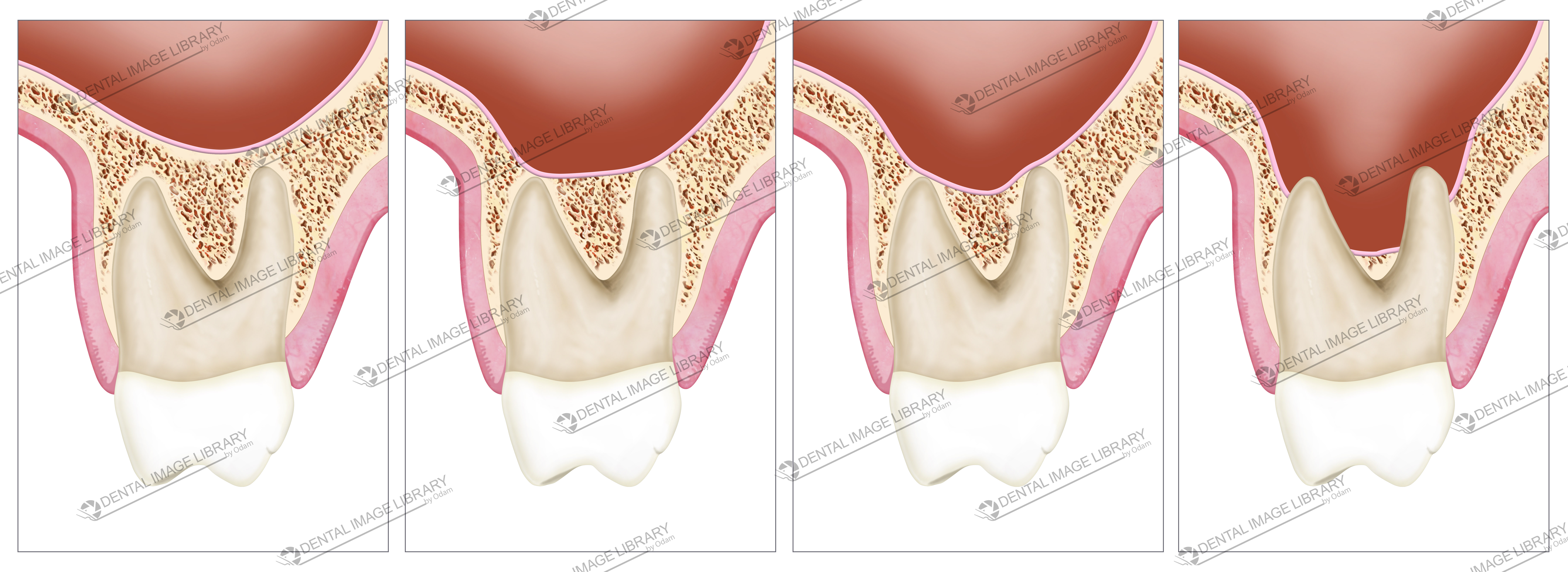
Relationship between maxillary molar roots and maxillary sinus floor.

Root-sinus classification.

The classification involves 4 types: ⠀
⠀
Type 0 = Root separate from the sinus floor⠀
Type 1 = Root in close contact with sinus floor⠀
Type 2 = Root projected laterally along the sinus cavity, but actually lateral or medial to it ⠀
Type 3 = Root projected into the sinus cavity⠀

Add to Wishlist loading
Product added! Browse Wishlist
The product is already in the wishlist! Browse Wishlist
Categories: Implantology, Oral Surgery, Periodontology Tags: classification, maxillary posterior teeth, maxillary sinus, maxillary sinus floor, molar, sinus lift
  • Description
  • Reviews (0)

Description

Relationship between maxillary molar roots and maxillary sinus floor.

Root-sinus classification.

The classification involves 4 types: ⠀
⠀
Type 0 = Root separate from the sinus floor⠀
Type 1 = Root in close contact with sinus floor⠀
Type 2 = Root projected laterally along the sinus cavity, but actually lateral or medial to it ⠀
Type 3 = Root projected into the sinus cavity⠀

White background.

8865 x 3236 pixels

150,11 x 54,8 cm (300 dpi)

JPEG

Reviews

There are no reviews yet.

Be the first to review “Root-sinus classification. 61JW00165” Cancel reply

Your email address will not be published. Required fields are marked *

Related products

  • Elisa Botton

    Tooth and implant attachment to surrounding tissue. 62JB00014

    45,00€
    Add to cart Show Details
  • Elisa Botton

    101JN00015

    45,00€
    Add to cart Show Details
  • Elisa Botton

    Representation of tooth and implant attachment to surrounding tissue. 62JB00007

    45,00€
    Add to cart Show Details
  • Elisa Botton

    Implant angulation and features of fenestration. 62JW00005

    40,00€
    Add to cart Show Details

TERMS AND CONDITIONS – LICENSING WITH THE DENTAL IMAGE LIBRARY

© Copyright 2023 - Dental Image Library
Elisa Botton - Via Mafalda di Savoia, 24/2. 45100 Rovigo - privacy@dentalimagelibrary.com - 3280657100 - bottonelisa@pec.it
  • Facebook
  • Instagram
Maxillary sinus anatomy. Frontal section of the nasal cavity and sinus Distal wedge procedure. Collection of 4 illustrations
Scroll to top