

Description
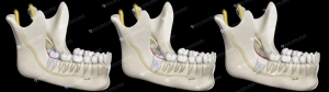
This medical illustration set shows upper airway obstruction during snoring compared with normal upper airway anatomy during breathing.
The first image shows a patent airway, with unobstructed airflow through the nasal cavity, soft palate and pharynx.
The second image illustrates airway obstruction caused by the posterior displacement of the tongue and soft palate, reducing airflow and leading to snoring.
This side-by-side comparison helps clearly visualize the anatomical changes involved in sleep-related breathing alterations.
Clinical relevance
Understanding the difference between a patent airway and airway obstruction is essential for the diagnosis and management of snoring and obstructive sleep conditions.
This illustration set is particularly useful for dental professionals, educators and medical communicators to explain airway dynamics, patient conditions and treatment approaches.
Ideal for lectures, patient education and scientific publications.
Technical details
- Number of illustrations: 2 (comparative set)
- Dimensions: 1741 × 1420 pixels (each image)
- Resolution: 300 DPI
- Format: JPG
- High-quality medical illustration suitable for digital and print use

 Elisa Botton
 Elisa Botton
 Elisa Botton
 Elisa Botton
Reviews
There are no reviews yet.